
Imagine you have a mysterious mucosal blistering disorder, and your entire mouth is lined with bleeding, open, devastatingly painful sores.
Now imagine that a medical provider is going to use a needle to inject lidocaine and cut pieces of flesh out of your already fragile, broken mouth in hopes of finding a diagnosis.
Sounds brutal, right?
Honestly – It’s not that bad, if you have the right kind of person performing the biopsies.
Oral biopsies – the right way!
To start, I was placed in a reclining position on the exam table with my head relaxed back on extra pillows. The doctor (a dermatologist) looked carefully at my mouth and identified the locations to collect meaningful samples. Then out came the tray of scary tools!
Truly, the only painful part of the process was the quick pinch and burn of the lidocaine going in through the needle. That was just a fleeting moment. If you can tolerate a vaccine or a blood draw, you can do this! I would actually describe it as more awkward than painful.
For about a minute after the injection, the doctor gently tested my sensation. Could I feel pressure? Could I feel pain? Once the pain was fully gone, she used a small, sharp tool to collect two shallow samples right along the margin of active lesions in my mouth. The whole cutting process was just a few seconds and I ever only felt a light touch. It was really no big deal at all!
After the samples were collected, she used several dental gauze rolls to stop bleeding – and I was done. No sutures needed! She prepared separate samples for H&E and DIF interpretation. It’s very important that BOTH of these types of biopsy are completed.
I went home and ate macaroni and cheese and ice cream. I didn’t even feel slight discomfort at the biopsy site the next morning. It was like it never happened.
But, sometimes biopsies go wrong
The successful biopsy wasn’t my first time at the rodeo. In January 2024, I had a poorly done biopsy. I had a long, difficult recovery and no meaningful answers when the pathology came back. I vowed to never put myself through such a painful and pointless procedure again.
For almost three weeks after the first biopsy, I couldn’t talk or eat without my mouth bleeding profusely. I couldn’t dry my face with a towel or brush my teeth. I couldn’t use a spoon or straw because the pressure on my sutured lip was too much – anything that touched the wound inevitably pulled more flesh away. The only way I got calories was by using a dropper to place liquid food into the back of my mouth. I lost a ton of weight, was constantly dizzy, and had constant low blood pressure. It took forever for the wound to heal. It was awful! I ended up having to use almost three weeks of FMLA leave because I was unable to work.
The sutures were supposed to be self-absorbing, but they never disintegrated at all. I had to endure going back for the extremely painful process of having the threads cut and dug out of my lip.
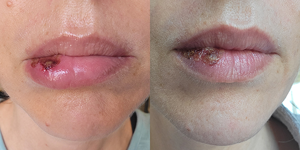
If you have a tolerance for gore, these photos are my healing at one and three weeks after the first biopsy. It took five to six weeks for this wound to heal to the point it didn’t bleed when touched.
Brave in the face of a second round of biopsies
Ten months after this negative experience and vowing never again, I changed my mind and had the biopsy described earlier in this post. I was so desperate to find a diagnosis and a biopsy just felt like the right path forward. It’s one of the bravest things I’ve done in the course of my illness.
I’m so glad I did it!
The second biopsy was nearly painless, healed within a day or two, never stopped me from being able to eat, and gave me the answers I had long been seeking.
I don’t have any photos of the healing process from my second biopsy because there was literally no issue to document.
What made these two procedures so different?
Biopsy 1 – The Bad One
- The provider conveyed confidence and competence, but was likely inexperienced
- The sample was not collected from a good location
- Sutures were poorly placed in my mouth
- Only a hematoxylin and eosin (H&E) biopsy was performed. This is insufficient as a diagnostic tool
- Samples were sent to a lab that may not have had enough experience to interpret the pathology
Biopsy 2 – The Good One
- The doctor had many years of experience collecting oral biopsies related to mucosal blistering diseases
- Multiple samples were gently collected from suitable locations
- No sutures were used, instead dental rolls were temporarily applied to absorb blood
- Both H&E and DIF (Direct Immunofluorescence) biopsies were completed, allowing a fuller diagnostic picture
- An experienced pathologist interpreted the samples
If you find yourself at a crossroads and think a biopsy might give you the answers you need… be brave – do it! But ask questions and make sure your healthcare provider is up to the task. It makes all the difference!
